Demystifying the Terminology: What We Call the Lower Part of the Leg
When we talk about the segment between your knee and your ankle, what exactly is the correct term? While commonly referred to as the "lower leg," this anatomical region has a more specific scientific name: the crus. However, for everyday understanding and most general discussions, "lower leg" is perfectly accurate and widely accepted. This crucial part of your body is more than just a simple connection; it's a complex structure supporting your entire upper body and facilitating virtually all forms of locomotion.
The lower part of the leg encompasses everything from the joint connecting it to the thigh (the knee) down to the joint connecting it to the foot (the ankle). It's a powerhouse of bones, muscles, nerves, and blood vessels, all working in concert to enable you to stand, walk, run, and jump. Understanding its various components and their functions is essential for anyone interested in human anatomy, fitness, injury prevention, or overall well-being.
The Core Structure: Bones of the Lower Part of the Leg
The foundation of the lower part of the leg is built upon two long bones: the tibia and the fibula. These bones provide the structural integrity, bear weight, and offer attachment points for the powerful muscles that control ankle and foot movement.
The Tibia (Shin Bone)
The tibia is the larger and more medial of the two lower leg bones, often referred to as the "shin bone." It's easily palpable along the front of your leg. As the primary weight-bearing bone, the tibia is incredibly robust. Proximally, it articulates with the femur to form the knee joint, transmitting the weight from your thigh down to your foot. Distally, it forms the medial malleolus, the prominent bony bump on the inside of your ankle, contributing significantly to the ankle joint's stability and structure.
The Fibula
Positioned laterally to the tibia, the fibula is a much thinner bone. While it plays a minimal role in weight bearing (only about 5-10% of body weight passes through it), its importance is immense for muscle attachment and stabilizing the ankle joint. Proximally, its head articulates with the tibia just below the knee, and distally, it extends further down to form the lateral malleolus, the bony protrusion on the outside of your ankle. This creates a protective arch for the ankle joint and is crucial for maintaining proper foot alignment and movement.
Together, the tibia and fibula create a stable framework that protects vital structures and allows for a wide range of movements at both the knee and ankle. Their precise arrangement and strong connective tissues ensure the integrity of the lower part of the leg under considerable stress.
Powering Movement: Muscles of the Lower Part of the Leg
The musculature of the lower part of the leg is complex and highly specialized, organized into distinct compartments by tough fascial sheets. These muscles are responsible for all movements of the foot and ankle, crucial for ambulation and balance. For a more detailed look at these intricate structures, you might find our article Lower Leg Muscles: Definition, Key Parts, and Full Anatomy incredibly insightful.
Anterior Compartment
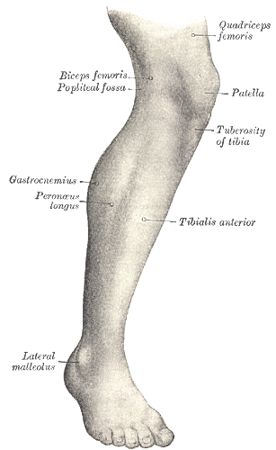
Located on the front of the shin, these muscles are primarily responsible for dorsiflexion – lifting the foot upwards towards the shin. The key player here is the tibialis anterior, essential for clearing the foot off the ground during walking and running, preventing trips and falls.
Lateral Compartment
Found on the outside of the lower leg, the peroneal (or fibularis) muscles dominate this compartment. Their main actions are eversion (turning the sole of the foot outwards) and assisting in plantarflexion (pointing the foot downwards). They play a critical role in ankle stability, especially on uneven surfaces.
Posterior Compartment (Superficial)
This is the largest and most well-known compartment, forming the bulk of the "calf." It's dominated by the gastrocnemius and soleus muscles, which together form the triceps surae. These powerful muscles are the primary movers for plantarflexion, enabling you to push off the ground when walking, run, jump, and stand on your toes. They merge to form the Achilles tendon, the strongest tendon in the body.
Posterior Compartment (Deep)
Lying beneath the superficial calf muscles, this compartment includes muscles like the tibialis posterior, flexor digitorum longus, and flexor hallucis longus. These muscles are crucial for inversion (turning the sole of the foot inwards) and fine motor control of the toes, supporting the arch of the foot, and assisting in plantarflexion.
The synergy between these muscle groups allows for the incredibly varied and precise movements that are fundamental to human locomotion and interaction with our environment.
Beyond Bones and Muscles: Nerves, Vessels, and Joints
While bones provide structure and muscles provide movement, the lower part of the leg also houses a sophisticated network of nerves and blood vessels, all converging at the ankle joint to ensure proper function and sensation.
Key Nerves
The major nerves of the lower leg originate from the sciatic nerve in the thigh. The common peroneal nerve wraps around the head of the fibula, dividing into superficial and deep branches that supply the lateral and anterior compartment muscles, respectively, and provide sensation to parts of the foot and lower leg. The tibial nerve descends through the posterior compartment, innervating the posterior muscles and providing sensation to the sole of the foot. Damage to these nerves can lead to significant motor and sensory deficits.
Major Blood Vessels
Blood supply to the lower leg is robust, primarily from the popliteal artery, which branches into the anterior tibial, posterior tibial, and peroneal arteries. These arteries ensure that all muscles, bones, and tissues receive a constant supply of oxygenated blood. Veins, such as the great and small saphenous veins, return deoxygenated blood to the heart, often prone to conditions like varicose veins due to gravity and long periods of standing.
The Ankle Joint
The ankle joint (talocrural joint) is formed by the distal ends of the tibia and fibula articulating with the talus bone of the foot. It's a hinge joint primarily responsible for dorsiflexion and plantarflexion. Its stability is paramount for walking and balance, provided by the bony architecture and strong ligaments that bind the bones together. The strength and flexibility of this joint, along with the subtalar joint (between the talus and calcaneus), are vital for adapting to uneven terrain.
The Vital Role of the Lower Part of the Leg in Daily Life
The lower part of the leg is a foundational segment of the human kinetic chain. It's not just a collection of anatomical parts; it's a dynamic system crucial for nearly every upright activity. From the simple act of standing to the complex mechanics of competitive sports, its contribution is profound.
This region acts as a shock absorber, cushioning the impact of ground forces during walking, running, and jumping. It’s also a powerful propulsive unit, generating the force needed to push off the ground. The muscles of the lower leg are constantly engaged, whether you're maintaining balance while standing still or performing explosive movements. Without healthy, functional lower legs, even basic mobility becomes challenging. For a deeper dive into how all the components of the lower limb, including the lower leg, are arranged and function together, exploring our article Exploring the Lower Limb: Topography and Muscle Arrangement can offer valuable insights.
Maintaining a Healthy Lower Leg: Tips and Considerations
Given its critical role, caring for your lower part of the leg is paramount. Neglect can lead to common and often painful conditions that impede daily activities.
- Strength Training: Regularly incorporate exercises that target both the anterior and posterior compartments. Calf raises (for gastrocnemius and soleus) are essential. Don't forget tibialis raises (lifting the front of the foot) to balance muscle development and prevent shin splints.
- Flexibility: Regular stretching, particularly of the calf muscles and Achilles tendon, can prevent tightness, improve range of motion, and reduce the risk of injuries like Achilles tendonitis.
- Proper Footwear: Choose shoes that provide adequate support, cushioning, and stability for your foot type and activity level. Worn-out shoes can alter gait mechanics and place undue stress on the lower leg.
- Gradual Progression: When starting new exercises or increasing intensity, do so gradually. Rapid increases in activity can overload the muscles and bones, leading to conditions like shin splints or stress fractures.
- Listen to Your Body: Pain is a signal. Don't push through persistent discomfort. Rest, ice, compression, and elevation (RICE) can often help with minor strains, but consult a healthcare professional for persistent or severe pain.
- Hydration and Nutrition: Adequate water intake and a balanced diet support muscle health, repair, and overall bodily function, including bone density.
By integrating these practices, you can significantly enhance the resilience and performance of your lower part of the leg, safeguarding your mobility and overall quality of life.
In conclusion, the lower part of the leg, whether you call it the crus, the shin, or the calf, is a remarkable feat of biomechanical engineering. Its intricate network of bones, muscles, nerves, and blood vessels enables the fundamental human abilities of standing, walking, and running. Appreciating its complexity and committing to its care through strength, flexibility, and smart lifestyle choices will ensure this vital segment continues to support your every move for years to come.