Understanding the Powerhouse: A Deep Dive into Lower Leg Muscles
The lower part of the leg, often overlooked in favor of more prominent muscle groups, is a complex and crucial anatomical region. Far from being just the "calf," it comprises an intricate network of muscles, tendons, and ligaments that enable a vast range of movements, from simple walking to complex athletic maneuvers. Understanding the definition, key parts, and full anatomy of the lower leg muscles is essential for athletes, fitness enthusiasts, healthcare professionals, and anyone interested in maintaining mobility and preventing injuries. These muscles are fundamental for ambulation, balance, and the powerful propulsion required in daily life and sports.
This comprehensive guide will explore the fascinating structure and function of the muscles that make up the lower part of the leg. We'll break down the major compartments, identify key muscles within each, discuss their roles, and offer insights into their functional importance and care.
Anatomy of the Lower Part Of The Leg: Structure and Compartments
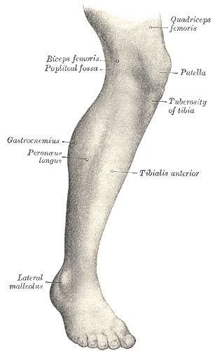
The lower leg, or crus, extends from the knee to the ankle. Its bony framework consists primarily of two long bones: the larger, weight-bearing tibia (shin bone) and the slender fibula, which provides muscle attachment sites and contributes to ankle stability. These bones are connected by an interosseous membrane, which also serves as an attachment point for several muscles and helps divide the lower leg into distinct compartments. This compartmental organization is a key anatomical feature, as it groups muscles with similar functions, nerve supply, and blood supply.
There are typically four main muscle compartments in the lower limb, each encased by tough fascial sheets:
- Anterior Compartment: Located on the front of the shin.
- Lateral Compartment: Positioned on the outside of the shin.
- Superficial Posterior Compartment: The large muscle mass at the back of the leg, commonly known as the calf.
- Deep Posterior Compartment: Lying beneath the superficial posterior muscles.
Each compartment houses specific muscles responsible for particular actions at the ankle and foot, contributing to the overall agility and strength of the lower part of the leg.
Key Muscle Groups of the Lower Part Of The Leg and Their Functions
Let's delve into the specific muscles found within each compartment and explore their vital roles.
The Anterior Compartment: Dorsiflexion and Toe Extension
The muscles in the anterior compartment are primarily responsible for dorsiflexion (lifting the foot towards the shin) and extending the toes. They are crucial for clearing the foot during the swing phase of walking and running.
- Tibialis Anterior: This is the largest muscle in this compartment, originating from the tibia and inserting into the medial cuneiform and first metatarsal. It's the primary dorsiflexor of the ankle and also assists with inversion of the foot (turning the sole inward). Weakness in this muscle can lead to "foot drop."
- Extensor Digitorum Longus (EDL): Originating from the tibia and fibula, this muscle divides into four tendons that extend the lateral four toes and assist in dorsiflexion.
- Extensor Hallucis Longus (EHL): Also arising from the fibula, its tendon extends the great toe (hallux) and aids in dorsiflexion.
- Fibularis (Peroneus) Tertius: A small muscle, often considered part of the EDL, that assists in dorsiflexion and eversion (turning the sole outward).
Practical Tip: Strengthening the tibialis anterior can help prevent shin splints, a common overuse injury, especially for runners.
The Lateral Compartment: Eversion and Plantarflexion
The lateral compartment muscles, often referred to as the peroneal muscles, are key players in eversion (turning the sole of the foot outward) and contribute to plantarflexion (pointing the foot downward). They are vital for ankle stability, particularly on uneven surfaces.
- Fibularis (Peroneus) Longus: Originating high on the fibula, its long tendon passes behind the lateral malleolus and under the foot to insert into the medial cuneiform and first metatarsal. It's a strong evertor and assists with plantarflexion.
- Fibularis (Peroneus) Brevis: Lying deeper than the longus, it originates lower on the fibula and inserts into the base of the fifth metatarsal. It also powerfully everts the foot and aids in plantarflexion.
Insight: These muscles are crucial for balance and preventing ankle sprains, especially lateral sprains, by stabilizing the ankle joint against excessive inversion.
The Posterior Compartment: The Calf Muscles and Beyond
The posterior compartment is the largest and most powerful, divided into superficial and deep layers. These muscles are predominantly responsible for plantarflexion, crucial for pushing off the ground during walking, running, and jumping.
Superficial Posterior Compartment: The Triceps Surae
This group forms the bulk of what we commonly call the "calf" and is collectively known as the triceps surae due to its three heads, all of which insert via the powerful Achilles tendon into the calcaneus (heel bone).
- Gastrocnemius: The most superficial calf muscle, it has two heads originating from above the knee joint. It's a powerful plantarflexor of the ankle and also assists with knee flexion. Its two heads give the calf its characteristic rounded shape.
- Soleus: Lying deep to the gastrocnemius, the soleus originates below the knee joint. It is a powerful plantarflexor, especially when the knee is bent, and is vital for endurance activities like standing and walking.
- Plantaris: A small muscle with a very long, slender tendon, it originates above the knee and inserts into the Achilles tendon or calcaneus. It's a weak assistant in plantarflexion and knee flexion, often considered vestigial.
Fact: The Achilles tendon, formed by the gastrocnemius and soleus, is the thickest and strongest tendon in the human body, capable of withstanding immense forces.
Deep Posterior Compartment: Fine Motor Control and Support
These deeper muscles provide support for the arch of the foot, fine-tune plantarflexion, and are involved in inversion and toe flexion.
- Tibialis Posterior: The deepest muscle, originating from the tibia, fibula, and interosseous membrane. Its tendon passes behind the medial malleolus and inserts into multiple tarsal and metatarsal bones. It's the primary invertor of the foot and a strong plantarflexor, crucial for maintaining the medial longitudinal arch of the foot.
- Flexor Digitorum Longus (FDL): Originating from the tibia, its tendon divides to flex the lateral four toes and assists in plantarflexion and inversion.
- Flexor Hallucis Longus (FHL): Arising from the fibula, its tendon flexes the great toe, crucial for gripping the ground, and aids in plantarflexion and inversion.
- Popliteus: A small, triangular muscle at the back of the knee that "unlocks" the knee joint from full extension by rotating the tibia medially (or femur laterally).
Actionable Advice: Proper care for the deep posterior muscles, especially the tibialis posterior, is vital for preventing conditions like flat feet and shin splints, often through targeted strengthening and stretching.
The Functional Importance of a Healthy Lower Part Of The Leg
The coordinated action of all these muscles is fundamental to human locomotion and stability. When you walk, the anterior compartment muscles initiate the swing phase by dorsiflexing the foot, preventing toe drag. As your foot makes contact with the ground, the posterior compartment muscles activate to control the landing and then powerfully plantarflex to push you forward. The lateral compartment muscles constantly work to maintain balance, adapting to uneven terrain and preventing inversion ankle sprains.
Beyond locomotion, strong and flexible lower leg muscles contribute to:
- Improved Athletic Performance: Enhanced jumping, sprinting, and agility.
- Balance and Stability: Reducing the risk of falls, especially in older adults.
- Injury Prevention: A balanced development of strength and flexibility across all compartments helps protect against common issues like Achilles tendonitis, calf strains, and chronic shin pain.
- Support for the Arches of the Foot: Crucial for shock absorption and efficient biomechanics.
Neglecting these muscles can lead to imbalances, pain, and a higher susceptibility to injuries. Regular stretching, targeted strengthening exercises, and choosing appropriate footwear are all key components of maintaining optimal lower leg health.
Conclusion
The lower part of the leg is a marvel of biomechanical engineering. Its intricate muscular structure, divided into distinct compartments, works in concert to enable everything from the most subtle foot adjustments to explosive athletic feats. From the dorsiflexors of the anterior compartment to the powerful plantarflexors of the posterior calf and the stabilizing evertors of the lateral compartment, each muscle plays a critical role. By understanding the definition, key parts, and full anatomy of these vital muscles, we gain a deeper appreciation for their functional importance and the steps necessary to keep them strong, flexible, and injury-free for a lifetime of active movement.