Unveiling the Complexity of the Lower Part Of The Leg: Topography and Muscle Arrangement
The human lower limb is a marvel of biomechanical engineering, designed to bear our weight, facilitate movement, and maintain balance. While often taken for granted, the intricate structure of the lower part of the leg—from just below the knee to the ankle—is a complex interplay of bones, muscles, tendons, nerves, and blood vessels. Understanding its topography and the precise arrangement of its muscles is crucial not only for healthcare professionals and athletes but for anyone seeking to optimize their physical well-being and prevent injuries. This comprehensive guide will explore the fascinating anatomy of this vital region, offering insights into its function, common issues, and practical tips for maintaining its health. The term "lower part of the leg" encompasses the area commonly referred to as the calf and the shin. It's a region defined by two main bones: the robust tibia (shin bone) and the slender fibula. These bones provide the framework, but it's the dynamic arrangement of muscles, organized into distinct fascial compartments, that truly empowers the leg with its remarkable range of motion and strength. To truly appreciate the foundational structure and common terminology for this area, you might find our article What Is The Lower Leg Called? Your Essential Anatomy Guide particularly insightful.Understanding the Anatomical Compartments of the Lower Leg
The muscles of the lower part of the leg are not just randomly distributed; they are meticulously organized into four distinct compartments by tough sheets of connective tissue called fascia. These compartments, each with its own group of muscles, nerves, and blood supply, allow for specialized functions while also protecting delicate structures. This compartmentalization is key to understanding both normal movement and certain medical conditions like compartment syndrome.The Anterior Compartment: Movers and Shapers of the Shin
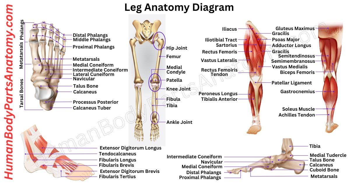
Positioned at the front of the tibia, the anterior compartment is primarily responsible for dorsiflexion (lifting the foot upwards towards the shin) and extension of the toes. This group of muscles is essential for clearing the foot off the ground during walking and running, preventing trips and falls.- Tibialis Anterior: This is the largest and most superficial muscle in this compartment, easily palpable along the shin bone. Its primary role is powerful dorsiflexion of the ankle and inversion of the foot (turning the sole inward). Weakness in this muscle can lead to "foot drop."
- Extensor Digitorum Longus: Originating from the fibula and tibia, this muscle extends the lateral four toes and also assists in dorsiflexion and eversion (turning the sole outward).
- Extensor Hallucis Longus: A deeper muscle that specifically extends the big toe (hallux) and also contributes to dorsiflexion and inversion.
- Fibularis (Peroneus) Tertius: Often considered part of the Extensor Digitorum Longus, this small muscle aids in dorsiflexion and eversion.
Clinical Insight: The anterior compartment is a common site for shin splints (medial tibial stress syndrome), an overuse injury characterized by pain along the shin bone, often due to repetitive impact activities without adequate conditioning or recovery. Strengthening the tibialis anterior through exercises like heel walks can help prevent this condition.
The Lateral Compartment: Stabilizers and Evertors
Located on the outer side of the fibula, the lateral compartment muscles are crucial for ankle stability and eversion of the foot. They help counteract the inward pull of the posterior muscles and play a significant role in adapting the foot to uneven surfaces.- Fibularis (Peroneus) Longus: A long, superficial muscle that runs down the lateral aspect of the leg. It powerfully everts the foot and assists in plantarflexion (pointing the toes downwards), contributing to the arch support of the foot.
- Fibularis (Peroneus) Brevis: Shorter and lying deep to the Longus, this muscle also everts the foot and assists in plantarflexion.
Practical Tip: Strengthening these muscles can significantly improve ankle stability, reducing the risk of inversion ankle sprains, which are among the most common sports injuries. Balance board exercises or resistance band eversion exercises are excellent for targeting this compartment.
The Posterior Compartment: Powerhouses of the Calf
The posterior compartment forms the bulk of the calf and is divided into superficial and deep groups. These muscles are primarily responsible for plantarflexion, crucial for pushing off the ground during walking, running, and jumping. Superficial Posterior Compartment: The Calf Muscles These are the most prominent muscles of the lower part of the leg, forming the characteristic bulge of the calf.- Gastrocnemius: The two-headed muscle that gives the calf its shape. It crosses both the knee and ankle joints, making it a powerful plantarflexor and also a knee flexor. It's highly active in explosive movements.
- Soleus: Located deep to the Gastrocnemius, the Soleus is a broad, flat muscle that only crosses the ankle joint. It's a crucial postural muscle, providing sustained plantarflexion, especially during prolonged standing or walking.
- Plantaris: A small, vestigial muscle with a very long tendon, sometimes absent. It weakly assists in plantarflexion and knee flexion.
Together, the Gastrocnemius and Soleus form the triceps surae, and their combined tendon forms the mighty Achilles tendon, the thickest and strongest tendon in the human body, connecting to the heel bone (calcaneus).
- Popliteus: A small, triangular muscle located behind the knee. It "unlocks" the extended knee, allowing flexion to begin, and also provides medial rotation of the tibia.
- Flexor Digitorum Longus: Flexes the lateral four toes and assists in plantarflexion and inversion.
- Flexor Hallucis Longus: Flexes the big toe (hallux) and strongly contributes to plantarflexion and inversion, playing a vital role in propelling the body forward during gait.
- Tibialis Posterior: Often called the "workhorse" of the leg, this muscle is a primary inverter and plantarflexor. It is fundamental for maintaining the medial longitudinal arch of the foot, crucial for shock absorption and efficient locomotion.
To dive deeper into the specific muscle definitions and their intricate anatomy, refer to our comprehensive guide on Lower Leg Muscles: Definition, Key Parts, and Full Anatomy.
Topographical Landmarks and Clinical Significance
Understanding the surface anatomy of the lower part of the leg allows for both self-assessment and clinical examination. Key palpable landmarks include: * The prominent anterior crest of the tibia (the shin bone itself). * The medial and lateral malleoli, the bony prominences on either side of the ankle, formed by the tibia and fibula respectively. * The robust Achilles tendon at the back of the heel. * The muscular bulk of the gastrocnemius in the calf. Beneath the surface, vital neurovascular structures navigate these compartments. The deep fibular nerve supplies the anterior compartment muscles, while the superficial fibular nerve supplies the lateral compartment. The tibial nerve and posterior tibial artery are housed within the deep posterior compartment. Damage to these nerves can lead to sensory deficits or muscle weakness, while vascular compromise can be limb-threatening. Conditions like compartment syndrome, where increased pressure within a fascial compartment compromises blood flow and nerve function, are medical emergencies requiring prompt attention. Recognizing symptoms such as severe pain disproportionate to the injury, swelling, tenderness, and tingling or numbness can be crucial for early diagnosis.Training and Maintaining a Healthy Lower Part Of The Leg
A strong, flexible, and well-balanced lower part of the leg is fundamental for overall mobility, athletic performance, and injury prevention. Incorporating a holistic approach to training this area is essential.- Strength Training:
- Calf Raises: Both straight-leg (targeting gastrocnemius) and bent-knee (targeting soleus) variations are crucial for plantarflexion strength.
- Tibialis Raises: Using resistance bands or specialized equipment to strengthen the anterior compartment muscles, preventing foot drop and shin splints.
- Ankle Eversion/Inversion: Resistance band exercises to strengthen the lateral and deep posterior compartment muscles, improving ankle stability.
- Flexibility: Regular stretching of the calf muscles (both gastrocnemius and soleus) is vital for maintaining ankle range of motion and preventing Achilles tendon issues. The anterior compartment muscles can also benefit from gentle stretching.
- Balance and Proprioception: Incorporate exercises like single-leg stands, wobble board exercises, and plyometrics to enhance the coordinated action of these muscles, improving stability and reactivity.
- Proper Footwear: Wearing supportive shoes that fit well and provide adequate cushioning can significantly reduce stress on the muscles and joints of the lower leg.
- Listen to Your Body: Overtraining is a common cause of lower leg injuries. Allow for adequate rest and recovery, and gradually increase the intensity and duration of your workouts.